tree in bud lesion
Without an obvious mass although a small central lesion is not excluded. Endobronchial tuberculosis the tree-in-bud lesions consist of either endo-luminal caseum or mixture of intra- and peri-bronchiolar caseation inflammatory cells and debris fibrosis and granuloma formation.

Tree In Bud Sign Marking Advancing Cavitary Tb A 4 Mm Coronal Ct Download Scientific Diagram
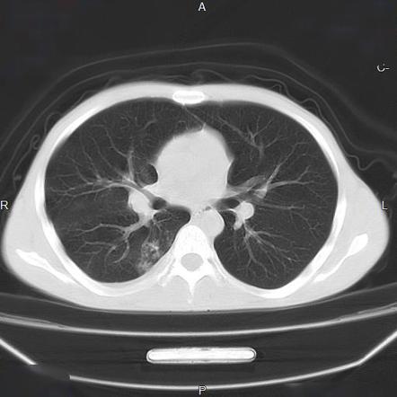
CT scan shows Tree in Bud lesions showing an appearance of multiple areas of centrilobular nodules with a linear branching pattern.

. Patterns of disease can provide clues to the most likely diagnosis. Earliest findings of endobronchial spread tuberculosis mostly from adjacent cavity is the formation of intrabronchiolar caseum. Tree in Bud appearance.
The nonclassic NTMB presents with chronic cough and as a bronchiectatic disease with centrilobular nodules and tree-in-bud pattern in relation to the bronchiectasis. Based on lesion localization and presence or suspicion of a concomitant bronchial disease for cats in this sample authors propose that the CT treeinbud pattern described in humans is also a characteristic of bronchial or bronchiolar plugging and bronchial disease in cats. The small nodules represent lesions involving the small airways.
The purpose of this study was to determine the relative frequency of causes of TIB opacities and identify patterns of disease associated with TIB opacities. Causes and imaging patterns of tree-in-bud opacities TIB opacities are most often a manifestation of infections or aspiration. In radiology the tree-in-bud sign is a finding on a CT scan that indicates some degree of airway obstruction.
High-resolution CT usually reveals small 24-mm centrilobular nodules and branching linear opacities of similar caliber originating from a single stalk Figs 2 3 4. Vascular abnormalities such as pulmonary tumor embolism can cause this sign. However to our knowledge the relative frequencies of the causes have not been evaluated.
The associated central bronchi are impacted. Patterns of disease can provide clues to the most likely diagnosis. Centrilobular nodules with a linear branching pattern are consistent with tree-in-bud appearance in a patient with endobronchial spreading of post-primary tuberculosis.
The list of the most frequent differential diagnoses for tree-in-bud sign includes infections with Mycobacterium tuberculosis nontuberculous mycobacteria and other bacterial fungal or viral pathogens. Non-infectious causes of the tree-in-bud sign include diffuse panbronchiolitis cystic fibrosis immotile cilia syndrome and congenital immunodeficiency states. Clusters of micronodules seen on 7-mm thick post-mortem radiographs with tuberculosis proved to be clusters of tree-in-bud lesions within the.
The tree-in-bud pattern occurs commonly in patients with endobronchial spread of Mycobacterium tuberculosis and is highly suggestive of active tuberculosis 2 3. Tree-in-bud sign refers to the condition in which small centrilobular nodules less than 10 mm in diameter are associated with centrilobular branching nodular structures 1 Fig. Generally these often result in bronchial wall thickening with replacement of the normally air-filled lumen with mucous or pus.
Slice thickness is 1 mm. The tree-in-bud pattern seen on CT represents radiologic sequelae of an infectious or in ammatory process. Bud measures 12 mm in diameter and is definitely bigger than parent bronchiole tree.
Figure 11 Non-tuberculous mycobacterial infection. The tree-in-bud sign is a nonspecific imaging finding that implies impaction within bronchioles the smallest airway passages in the lung. Multiple causes for tree-in-bud TIB opacities have been reported.
As a result involved bronchioles are more conspicuous on computed tomography imaging. However vascular lesions involving the arterioles and capillaries may simulate the centrilobular small nodules and. Hi doctor My CT scan says defined streaky opacity with associated loss volume and clustered tree in bud nodules have developed in the anterior segment of the upper left lobe.
TIB opacities are most often a manifestation of infections or aspiration. The tree-in-bud sign is a commonfinding in HRCT scans. We investigated the pathological basis of the tree-in-bud lesion by reviewing the pathological specimens of bronchograms of normal lungs and contract radiographs of the post-mortem lungs manifesting active pulmonary.
PV pulmonary vein. Rare differential diagnoses are malignant conditions. Lymph node involvement at the carina level were noted.
Red bud lesions 766953 Asked August 11 2021 353 PM EDT A month ago a ref bud tree planted in my garden a year ago started showing 1-2 mm white sticky spots on the trunk. The tree-in-bud sign indicates bronchiolar luminal impaction with mucus pus or fluid causing normally invisible peripheral airways to become visible 80. Generally these often result in bronchial wall thickening with replacement of the.
We investigated the pathological basis of the tree-in-bud lesion by reviewing the pathological specimens of bronchograms of normal lungs and contract radiographs of the post-mortem lungs manifesting active pulmonary tuberculosis. It is not specific for a single disease entity but is a direct sign of various diseases of the peripheral airways and an indirect sign of bronchiolar diseases such as air trapping or sub-segmental consolidation. Histopathology The tree-in-bud pattern seen on CT represents radiologic sequelae of an infectious or inflammatory process.
There are no criteria for invasion of the aorta or the bronchial tree. Post-mortem radiograph of patient with active pulmonary tuberculosis demonstrating tree-in-bud lesion boxed area with smooth marginated bronchiole tree and distal clubbed end bud. The patient had an oesophageal lesion below the carina extending longitudinally 6 cm.
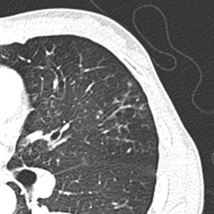
The tree-in-bud-pattern of images on thin-section lung CT is defined by centrilobular branching structures that resemble a budding tree. The tree-in-bud-pattern of images on thin-section lung CT is defined by centrilobular branching structures that resemble a budding tree. The remaining pulmonary parenchyma demonstrated scattered tree-in-bud pattern with lower lobe predominance and without pleural effusion.
Volume59 Issue1 JanuaryFebruary 2018 Pages32-42 Related Information. Cavitation and mediastinal lymphadenopathy are rare in nonclassic NTMB 18. This finding is considered classical for endobronchial TB as in this case.
The impression at the end said a focus of bronchitis and bronchiolitis. However has been described in other conditions as well.

Kjr Korean Journal Of Radiology

Cylindrical Bronchiectasis And Tree In Bud Pattern In Lower Lobes And Download Scientific Diagram

Tree In Bud Sign Lung Radiology Reference Article Radiopaedia Org
Tree In Bud Sign Lung Radiology Reference Article Radiopaedia Org

Co Rads 2 With Tree In Bud Sign A 27 Year Old Male Attended The Download Scientific Diagram

Pdf Tree In Bud
Cylindrical Bronchiectasis And Tree In Bud Pattern In Lower Lobes And Download Scientific Diagram
View Of Tree In Bud The Southwest Respiratory And Critical Care Chronicles

References In Causes And Imaging Patterns Of Tree In Bud Opacities Chest

Tree In Bud Pattern Pulmonary Tb Eurorad

Tree In Bud Pattern Pulmonary Tb Eurorad

Computed Tomography Of The Chest Showed Nodular Opacities With Tree In Download Scientific Diagram
2
Tree In Bud Sign Lung Radiology Reference Article Radiopaedia Org
2
Tree In Bud Sign Lung Radiology Reference Article Radiopaedia Org
View Of Tree In Bud The Southwest Respiratory And Critical Care Chronicles

Tree In Bud Sign Lung Radiology Reference Article Radiopaedia Org

Areas Showing A Mosaic Pattern Of Attenuation And Tree In Bud Opacities Download Scientific Diagram